Introduction
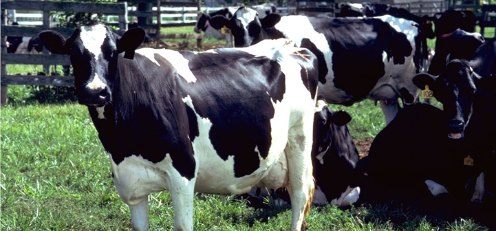
The periparturient period is especially important for the health of dairy cows. Among health problems occurring during this period is an accumulation of excessive interstitial fluid in extravascular spaces of the udder and adjacent tissues of some animals. Occurrence of udder edema varies greatly in different herds, with incidences ranging from 18 to 96% of all cows observed at calving (Al-Ani and Vestweber, 1986). Although the swelling usually diminishes after parturition without special care, management problems and permanent damage are likely (Vestweber and Al-Ani, 1983).
Please check this link first if you are interested in organic or specialty dairy production.
Physiology of Udder Edema
Intravascular and interstitial fluids
Over a century ago, Starling (1896) described the basic principles regulating movement of fluids between intravascular circulation and adjacent tissues. During the intervening 102 years, it has been established that relative concentrations of solute in intracellular and interstitial fluids markedly influence colloidal pressure, fluid retention, and edema (Mobarhan, 1988). Movement of fluid between blood vessels and interstitial fluid is controlled by a balance between hydrostatic and osmotic pressures (Staub, 1978). When both pressure and premeability are normal, the endothelial barrier restricts transfer of both fluids and solutes. Starling’s (1896) equation as interpreted by Vestweber and Al-Ani (1983, 1984) shows fluid transfer from intravascular to interstitial spaces is controlled by changes in either hydrostatic pressure or vascular permeability.
Basic principles of edema formation have been reviewed by Vestweber and Al-Ani (1983). Decreased plasma colloidal pressure, increased capillary blood pressure, obstruction of lymphatic drainage, and retention of sodium and water were listed as basic causes of generalized edema. When increased capillary pressure or obstruction of lymphatic drainage are localized, edema may also be localized as in udder edema.
Hydrostatic pressure
Greater incidence and persistence of udder edema in first-calf heifers than in multiparous cows may result from the less well developed vascular circulation in heifers (Emery et al., 1969). Vascular changes occur rather abruptly during latter stages of a heifer’s first pregnancy. Mammary blood flow increases at least three-fold during the two weeks before calving (Al-Ani and Vestweber, 1986). Vestweber and Al-Ani (1985) compared milk vein blood pressures in eight cows with udder edema and three control cows. Venous blood pressure increased between two weeks prepartum and calving by approximately 35% in edematous cows but remained constant in cows without edema. At parturition, mammary blood flow of edematous cows averaged only 85% that of control cows. It seems reasonable that elevated arterial blood flow to the udder in association with decreased venous blood flow from the udder would elevate capillary hydrostatic pressure.
Osmotic pressure
Osmotic pressure resulting from the higher protein content of blood serum relative to interstitial fluid (Vestweber and Al-Ani, 1984) opposes hydrostatic pressure. Increased hydrostatic pressure without a corresponding change in osmotic pressure could increase interstitial fluid volume if additional fluid is not removed by the lymph system. In addition, increased permeability of the endothelial barrier resulting from some injury would render it less restrictive to both fluid and proteins. Decreased osmotic pressure due to a decrease in protein concentration in blood serum relative to interstitial fluid could allow hydrostatic pressure to move fluid into tissues. If accumulation of edematous fluid is determined by hydrostatic and osmotic pressures, increases in either pressure or vascular permeability could result in edema (Vestweber and Al-Ani, 1983, 1984).
Increased vascular permeability is considered more likely than increased hydrostatic pressure to elevate protein in interstitial fluid relative to serum (Staub, 1974). Therefore, relatively high serum to interstitial fluid ratios of protein in edematous cows has been considered as evidence for a lesser role for change in vascular permeability in development of udder edema (Vestweber and Al-Ani, 1984). Declines in serum protein between two weeks prepartum and calving were similar for nine edematous and three non-edematous cows. Unfortunately, interstitial fluid for comparison with serum is available only for edematous cows.
Causes of edema
Although the exact cause of udder edema has not been identified, a number of physiological and management factors have been associated with its occurrence. Predisposition to edema may be inherited, decrease with increasing parity, and be more likely in older first-calf heifers. Excess prepartum intakes of energy, sodium, or potassium may also increase likelihood of edema. Research relative to these factors has been reviewed extensively (Al-Ani and Vestweber, 1986; Vestweber and Al-Ani, 1983) and will not be repeated here. Rather, we will propose an alternative hypothesis relating udder edema to oxidative stress.
Oxidative Stress
Partial reduction of oxygen during metabolism
Although oxygen is essential for aerobic life, it can be toxic under certain conditions. Animals do not use energy in feed directly for maintenance and productive purposes but first convert it into adenosine triphosphate (ATP) which releases energy as needed. The process has been compared with deposits into or withdrawals from a bank with ATP serving as the energy currency (Gutteridge and Halliwell, 1994). The ATP is produced by oxidative phosphorylation, a process driven by electrons derived from metabolism of feed. Energy released from these electrons as they are transferred down a chain of enzymes in animal cells is used to generate ATP. At the end of the chain, spent electrons are combined with oxygen to form its completely reduced metabolite, water. Air-breathing animals have achieved great energetic efficiency by using oxygen to accept electrons in this manner.
Problems may arise when oxygen is not completely reduced to water. It has been estimated that 2 to 5% of the electrons may escape from intermediate carriers in the chain and partially reduce oxygen to a free radical, superoxide (Levine and Kidd, 1985). Other sources of superoxide (O2–) include mechanisms which use oxygen to detoxify foreign substances in the body, other oxidative enzymes, and spontaneous nonenzymatic oxidation of biological molecules. Anything which increases metabolic rate or activity of oxidative enzymes could increase the number of electrons transferred and amount of oxygen consumed. Rapid growth, high milk production, or excessive exposure to aflatoxins, for example, could thus elevate generation of O2–. Superoxide is reduced further to hydrogen peroxide (HOOH). Both O2– and HOOH are unavoidable products of normal metabolic processes and are not always harmful if metabolized properly. Neither O2– nor HOOH are sufficiently reactive by themselves to initiate peroxidative chains, so damage by them is believed to result from their conversion into more reactive free radicals. It is in this conversion that catalytic transition elements, especially Fe, are believed to have harmful effects on oxidative metabolism (Gutteridge and Halliwell, 1994).
Role of iron in oxidative problems
The ability of Fe to transfer single electrons in controlled biological oxidations is essential for life as we know it, but it also gives Fe a pivotal role in harmful oxidative reactions. Iron normally is safely complexed in specific molecules which keep it away from the initial partially reduced oxygen metabolites, O2– and HOOH. Release of catalytic Fe becomes more likely under conditions of dietary imbalance, trauma, or stress often accompanying calving (Madsen et al., 1990). Saturation of safe binding sites (Gutteridge and Quinlan, 1993) by excessive Fe intake may also contribute to the problem. When catalytic Fe comes in close proximity with O2– and HOOH, O2– reduces Fe3+ to Fe2+ which splits into a hydroxyl ion and an extremely reactive hydroxy radical (∃OH).
Generation of ∃OH causes two distinct types of damage depending on location (Gutteridge and Haliwell, 1994). When ∃OH is produced in blood plasma or elsewhere in the “free pool,” it may become the focal point of peroxidative chains which can damage cellular and subcellular membranes. In contrast, catalytic Fe nonspecifically associated with an important molecule, such as an enzyme, may catalyze ∃OH which damages the molecule at the site where iron is located.
Defense against reactive forms of oxygen
Fortunately, animals possess a remarkably efficient antioxidant system which protects them against reactive oxygen metabolites and their toxic products resulting directly or indirectly from catalytic Fe (Gutteridge and Halliwell, 1994). First, O2– and HOOH are converted into forms which do not react with catalytic Fe by superoxide dismutase and glutathione peroxidase. Second, Fe is oxidized to less reactive forms by ceruloplasmin and complexed safely by transferrin and lactoferrin to prevent potential reactions with O2– and HOOH which could produce ∃OH. Third, important molecules may be protected against catalytic iron by Zn2+ which has an outer electron makeup similar to Fe2+ but is not involved in electron transfer (Bray and Bettger, 1990; Oteiza et al., 1995). Nothing is perfect, so despite the protective systems described above, some O2– and HOOH may escape enzymatic control, and some catalytic Fe may exist to generate ∃OH.
A fourth level of protection consists of molecules which terminate chain reactions. Protein, primarily albumin, may account for up to half of the total peroxyl radical-quenching capacity of blood plasma (Wagner et al., 1987). Other radical quenchers include lipid soluble α-tocopherol, β-carotene, and retanoic acid and water soluble ascorbate, phenolics, and urate (Gutteridge and Halliwell, 1994; Machlin and Bendich, 1987). Fifth, cytotoxic aldehydes produced when peroxidized lipids decompose are degraded by aldehyde dehydrogenases. An example is xanthine oxidase which also helps keep Fe in the less reactive ferric form (Emery, 1991), thereby conserving reducing equivalents, α-tocopherol, and other chain-breaking antioxidants.
Source of antioxidants
Endogenous production of transferrin, lactoferrin, ceruloplasmin, serum albumin, antioxidant enzymes, and glutathione is dependent on absorbed amino acids. An adequate supply of amino acids is dependent, in turn, on adequate amounts and appropriate balance of rumen degradable (nitrogen and sulfur) and escape protein with energy. Also of endogenous origin are ubiquonone and ascorbate for which the diet must supply fibrous and nonfibrous carbohydrates in adequate amounts and balance. The diet must supply vitamin A for immune function, α-tocopherol (vitamin E), and β-carotene as chain-breaking antioxidants, Cu, Zn, and Mn for superoxide dismutase, Se for glutathione peroxidase, Fe for catalase, Fe and Mo for xanthine dehydrogenase, Zn to displace catalytic Fe, and Mg and Zn to stabilize membranes and maintain cellular integrity.
Oxidative stress and disease
Pitzen (1993) has employed the prooxidant-to-antioxidant ratio (PAR) to minimize oxidative stress and related problems in dairy cows. The PAR increases when prooxidants such as Fe exceed antioxidants and cofactors such as vitamin E, β-carotene, Cu, Zn, Se, and Mn. Pitzen’s (1993) approach is to lower prooxidant intake when possible, but when this is difficult, such as is often the case with Fe, it may be necessary to increase antioxidant concentrations to counteract the excessive prooxidant.
Involvement of oxidative stress in etiologies of certain disorders of dairy cattle is suggested by reductions in incidence of retained placenta (Harrison et al., 1984) and mastitis (Smith et al., 1984; Weiss et al., 1997) when the antioxidant nutrients vitamin E and Se are supplemented. Analysis of records from more than 61,000 cows suggests that udder edema may share common causes with retained placenta and mastitis (Gröhn et al., 1989). Our hypothesis is that oxidative stress could increase severity of udder edema either directly by damage to membranes or indirectly by altering steroidogenesis.
Possible Association between Oxidative Stress and Udder Edema
Oxidative stress and membrane damage
Reports that concentrations of protein and globulin in blood fall as cows near parturition (Larson and Hays, 1958; Larson and Kendall, 1957; Vestweber and Al-Ani, 1984) suggest the possibility that vascular permeability to proteins may have increased. Intravenous infusion of plasma proteins was effective in treating udder edema (Larson and Hays, 1958). These observations led Mueller et al. (1989) to investigate effectiveness of vitamin E, an important antioxidant in membranes, in reducing severity of udder edema in primigravid heifers. Forty heifers were fed fescue and orchard grass hay free choice plus 8 lb/day of commercial dairy concentrate. Beginning six weeks before expected parturition and continuing daily until calving, half of the heifers were given 1,000 IU of vitamin E as d-α-tocopheryl acetate by gelatin capsule. The remaining heifers served as a control. Concentrations of globulin and total proteins in serum were measured using materials and methods in commercial kits (Total Protein No. 540 and Total Globulin No. 560, Sigma Chemical Co., St. Louis, MO).
Calves remained with their dams for 24 hours. At the PM milking on 1, 3, 7, and 14 days after calves were removed, milk yield, edema score, and udder measurements were recorded. Each heifer was scored for edema prior to milking using a scale of 0 (no edema) to 4 (very severe edema) by a single evaluator. Udder floor areas were measured, before and after the PM milking to objectively quantify severity of edema. Sheets of paper were pressed against wet teat ends before and after milking (Seykora and McDaniel, 1986). Area of the quadrilateral defined by the four spots was determined by connecting two spots with a diagonal line from which perpendicular lines were drawn to each of the remaining two spots (Figure 1). The sum of the areas of the resulting four right triangles was then calculated. Percentage decrease in udder floor area after milking was then determined. It was assumed that the more rigid and edematous the udder, the less udder floor area would decrease with removal of milk.

Udder edema, as evaluated by less decrease in udder floor area after milking, tended to be less severe for vitamin E supplemented heifers than for controls (Table 1) during the first three measurements. Differences were statistically significant on day 3 (day 4 of lactation). Milk yields corresponding to udder measurements were higher for vitamin E supplemented compared with unsupplemented heifers on the first 3 days and tended to be higher on day 7 and 14. Daily milk yield for vitamin E supplemented versus unsupplemented heifers averaged 56.3 versus 46.4 lb (P < 0.01) through the first 12 weeks of lactation. This suggests that incomplete evacuation of edematous udders during the critical period of early lactation could be reflected in lower milk yield throughout lactation.
| Day1 | Udder shrink | Milk yield | ||||
|---|---|---|---|---|---|---|
| Control | Vitamin E | P > F | Control | Vitamin E | P > F | |
| ————– (%) ————– | ————– (kg) ————– | |||||
| 1 | 14.7 | 22.9 | 0.10 | 3.6 | 6.9 | 0.02 |
| 2 | 19.0 | 24.5 | 0.16 | 4.7 | 8.4 | 0.01 |
| 3 | 16.0 | 24.4 | 0.02 | 6.0 | 8.2 | 0.01 |
| 7 | 24.4 | 26.6 | 0.57 | 7.8 | 9.7 | 0.10 |
| 14 | 38.4 | 39.2 | 0.86 | 10.3 | 12.3 | 0.08 |
| 1Day plus 1 equals day of lactation. | ||||||
Serum total protein and globulin concentrations at parturition were lower in 14 heifers with mild to severe edema than in 26 heifers without edema (Table 2). This contrasts with an earlier comparison (Vestweber and Al-Ani, 1984) between nine edematous and three nonedematous cows in which these measurements did not differ. Lower concentrations of protein in serum of edematous heifers is in harmony with increased vascular permeability allowing protein to leak into interstitial spaces. This could reduce osmotic forces which oppose intravascular hydrostatic pressure and allow edematous fluid to accumulate.
| Udder edema | |||
|---|---|---|---|
| No | Yes | P > F | |
| Number of heifers | 26 | 14 | |
| Total protein (g/dl) | 6.58 | 6.11 | 0.001 |
| Total globulin (g/dl) | 3.20 | 3.03 | 0.03 |
Udder edema generally appears between two weeks prepartum and calving during which period blood flow to the mammary gland increases three-fold (Al-Ani and Vestweber, 1986). We suggest the resulting increase in oxygen supply to the rapidly developing mammary gland over such a short period could produce a condition analagous to ischemia-reperfusion injury. Ischemia-reperfusion injury accompanies restoration of blood flow to a tissue following a reduction or blockage (Simpson et al., 1987). Tissue damage results not so much from oxygen deprivation during the blockage as from free-radical reactions caused by the surge of oxygenated blood following restoration of circulation. Neutrophils infiltrate tissues and, with xanthine oxidase stimulated by increased oxygen, produce O2– and HOOH from which the potent inducers of lipid peroxidation, #OH radicals, are produced (Simpson et al., 1987). Peroxidation of polyunsaturated fatty acids in membranes compromises their integrity. A similar reaction accompanying increased blood flow to the mammary gland of heifers nearing parturition may contribute to edema.
Oxidative stress and altered steroidogenesis
All steroid hormones are derived from cholesterol by modifications produced by enzymes dependent on cytochrome P-450. Susceptibility of these steroidogenic enzymes to lipid peroxidation (Staats et al., 1988; Takayanagi et al., 1986) could alter synthesis of steroid hormones under oxidative stress. Hydroxylases specific to cytochrome P-450 appear to differ in their vulnerability to free radical attack. When adrenal microsomes were depleted of vitamin E in vitro, 17α-hydroxylase and 17,20-lyase were among the enzymes most sensitive to inactivation (Takayanagi et al., 1986). Fetal adrenal cortisol, which increases markedly preceding parturition (Casey et al., 1985), acts on the placenta to increase activities of 17α-hydroxylase and 17,20-lyase. The 17α-hydroxylase is necessary for cortisol synthesis, and 17α-hydroxylase and 17,20-lyase are required in both pathways leading to 17β-estradiol.
Although speculative at this point, unequal vulnerability of specific steroidogenic enzymes to oxidative stress could contribute to udder edema (Miller et al., 1993). Steroidogenesis proceeds by different pathways and inadequacy of a key enzyme for one pathway may misdirect the reaction (Bhagavan, 1978). For example, if 17α-hydroxylase and 17,20-lyase are damaged more than 21-hydroxylase (Takayanagi, 1986), adrenal lipid peroxidation can be more inhibitory of cortisol, androgen, and estrogen than of mineralocorticoid synthesis. A defect in synthesis of cortisol with the resultant increase in adrenocorticotropic hormone (ACTH) could also provoke overproduction of corticosterone and 11-deoxycorticosterone (Yanase et al., 1991). This could cause Na retention and expansion of blood volume.
Individuals afflicted with a recognized congenital deficiency of 17α-hydroxylase and 17,20-lyase become edematous but fail to develop sexually (Yanase et al., 1991). It seems reasonable that relative impairment of these enzymes by oxidative stress could contribute to formation of udder edema. In this connection, it is interesting that udder edema score was negatively correlated with plasma 17β-estradiol (Malven et al., 1983). Excess dietary K could aggravate the edema via the renin-angiotensin system by increased K excretion accompanied by Na and water retention (Sanders and Sanders, 1981).
We tested our hypothesis that oxidative stress may contribute to udder edema via alteration of steroidogenesis in a comparison with 38 periparturient heifers. The heifers were divided into two groups with udder edema either more severe or less severe than the average of all heifers on the basis of udder floor measurements as described previously. Blood plasma sampled at calving was assayed for antioxidant activity (Glazer, 1988), progesterone, 17β-estradiol, and corticosterone (Diagnostic Products Corp., Los Angeles, CA).
Relationships among udder edema, plasma antioxidant status, corticosterone, 17 β-estradiol (E2), and progesterone (P4) were evaluated using odds ratios (Fletcher et al., 1988). Heifers with above-average plasma antioxidant status were only 21% as likely as heifers below average in this measurement to develop above-average udder edema (Table 3). Antioxidant status was not related to corticosterone, E2, or P4 in plasma. Ratios of corticosterone to E2 or P4 were then calculated individually for each heifer. Antioxidant status and corticosterone to P4 ratio were not related. However, heifers with above-average compared with below-average antioxidant status were only 16% as likely to have an above-average corticosterone to E2 ratio. Heifers with an above-average corticosterone to E2 ratio were almost four times more likely to have udder edema (Table 3). It should be remembered that E2, but not corticosterone or P4, is dependent on 17α-hydroxylase and 17,20-lyase (Yanase et al., 1991), two enzymes among the ones most vulnerable to oxidative stress (Takayanagi et al., 1986). These results, therefore, provide indirect evidence in support of the concept that oxidative stress may contribute to udder edema through selective damage to steroidogenic enzymes.
| Plasma antioxidants | Udder edema | |
|---|---|---|
| Udder edema | 0.21b | —- |
| Corticosterone (C)c | 1.12 | 2.14 |
| Estradiol (E2)d | 1.11 | 0.42 |
| Progesterone (P4)c | 1.20 | 1.37 |
| C/E2 ratio | 0.16b | 3.89b |
| C/P4 ratio | 0.64 | 1.62 |
|
aIf statistically significant, the relationship denoted by an odds ratio is positive if >1.0 and negative if <1.0. bP < 0.05. cSynthesized independently of 17α-hydroxylase or 17,20-lyase. dSynthesis dependent on 17α-hydroxylase and 17,20-lyase. |
||
Vitamin E and Zn as antioxidants
Fifty-six primigravid heifers, managed as described previously, were divided into seven blocks of eight animals according to expected calving date. Eight treatments were assigned randomly within each block and given daily by gelatin capsule during the last six weeks prepartum. Treatments included all combinations of vitamin E, Zn, and Fe in a 2x2x2 factorial arrangement. Daily amounts of supplements were 1,000 IU vitamin E from 2 g of d,lα-tocopheryl acetate; 800 mg of Zn, half from 4 g Zn methionine and half from 1.13 g ZnSO4; and 12 g Fe from 60 g FeSO4•7H2O. For 1,100-lb heifers consuming 1.8% of body weight, these amounts of trace minerals would provide 90 ppm of Zn and 1,300 ppm of Fe in dietary DM intake.
Decrease in udder floor area was measured as described previously. Blood plasma collected at calving was assayed for α-tocopherol (Hidiroglou, 1989) and unsaturated iron-binding capacity (UIBC) using materials and methods from a commercial kit (Sigma Diagnostics, St. Louis, MO).
Heifer groups were compared according to whether or not vitamin E or Zn was supplemented in the presence or absence of excess Fe (Figure 2). Severity of edema in each heifer was determined in relation to the average within a comparison. It was assumed a rigid, edematous udder would decrease in size after removal of milk less than a nonedematous udder. Smaller decreases in udder floor area thus represent increasing severity of udder edema. Strengths of associations among prooxidants (Fe), antioxidants (Zn and vitamin E), plasma α-tocopheral, UIBC, and udder edema were calculated as odds ratios (Fletcher et al., 1988). Statistically significant odds ratios denote positive relationships when greater than one and negative relationships when less than one. Take, for example, two groups of heifers, one supplemented and the other unsupplemented with vitamin E. Within each group are heifers with udder edema more severe or less severe than the average. An odds ratio of 0.22 calculated from numbers of heifers in the four subgroups indicates supplemented heifers are only 22% as likely as unsupplemented heifers to have udder edema more severe than average.
When Fe was not excessive, heifers supplemented with vitamin E were only 19% as likely as unsupplemented heifers to have edema more severe than average (Figure 2). Supplemental Zn did not appear to reduce severity of udder edema when Fe was not excessive. In contrast, when Fe was excessive, vitamin E was ineffective in reducing severity of edema, but heifers supplemented with Zn were only 17% as likely to have greater-than-average severity of udder edema as unsupplemented heifers. When effects of supplemental vitamin E or Zn were compared regardless of Fe intake, severity of udder edema was reduced by vitamin E but not by Zn.

Implications
Research reviewed by Al-Ani and Vestweber (1986) and Vestweber and Al-Ani (1983) suggests management practices to minimize udder edema include limiting prepartum intakes of salt and high-potassium feeds and avoiding excessive prepartum conditioning. Severity of udder edema may also be increased by prooxidants such as excess Fe but decreased by antioxidants such as vitamin E and Zn. Modern management practices may expose dairy cows to excessive proxidants, so antioxidant requirements may be higher than generally recognized. Intakes of antioxidant nutrients sufficient to control prooxidant/antioxidant balance effectively may exceed amounts contained in average feedstuffs. Supplementation with all known nutrients required for antioxidant defense in adequate and balanced amounts would therefore be beneficial.
Protein and energy in optimum amounts to meet amino acid requirements for manufacture of Fe complexing agents and other endogenously produced antioxidants is of basic importance. Potential prooxidants including aflatoxins, pesticides, products of heat-damaged feed, and excessive Fe and Mo should be avoided as much as possible. Finally, mineral and vitamin supplements are needed. Based on present knowledge for dairy cows, and under average conditions, 0.3 ppm Se, 20 ppm Cu, 60 ppm each of Zn and Mn, and 0.25% Mg in dietary DM plus at least 1,000 IU/day of vitamin E appear to be adequate. Additional vitamin E may partially compensate for inadequacies of other antioxidants under some conditions, but cost effectiveness should be considered. Cellular oxidation/reduction balance may vary depending on environment, diet, disease, and other factors, so it may be necessary to adjust amounts of these nutrients accordingly.
Summary
Udder edema is caused by excessive accumulation of fluid in extravascular spaces of the udder and surrounding tissues. Usual recommendations for minimizing the problem include avoidance of excessive salt, potassium, or body condition before calving. Increasing evidence suggests oxidative stress may also contribute to udder edema. Oxidative stress results when partially reduced metabolites of oxygen produced during normal metabolism and increased by prooxidants, such as excess dietary Fe, exceed the cow’s antioxidant defense mechanisms. When reactive oxygen metabolites are not safely controlled, lipid peroxidation, damage to critical molecules, and ultimately disease conditions may result. Possible changes relative to udder edema include injury to membrane integrity or damage to specific steroidogenic enzymes, thereby altering synthesis of steroid hormones. Avoidance of potential prooxidants when possible and supplementation with all known nutrients required for antioxidant defense are thus important. Vitamin E, vitamin A, and β-carotene must be supplied by the diet, whereas vitamin C and glutathione may be of endogenous or dietary origin. Most other antioxidant molecules are produced endogenously, but dietary nutrients essential for their manufacture include protein (N and S), energy, Cu, Zn, Mn, Se, and Mg. The Fe and Mo are also essential but can be prooxidants when present in excess amounts.
Acknowledgment
Appreciation is expressed to Zihpro Corporaton (Eden Prairie, MN) and BASF Corp. (Mt. Olive, NJ) for supporting parts of our research.
Author Information
F. J. Mueller
Kalmbach Feeds Inc., Upper Sandusky, OH
J. K. Miller, M. H. Campbell
Department of Animal Science
The University of Tennessee, Knoxville
F. C. Madsen
Suidae Technology, Greensburg, IN
References
Al-Ani, F.K., and J.G.E. Vestweber. 1986. Udder edema: An updated review. Vet. Bull. 56:763.
Bhagavan, N.W. 1978. Biochemistry, 2nd ed. J. B. Lippincott Co., Philadelphia, Pa.
Bray, T.M., and W.J. Bettger. 1990. The physiological role of zinc as an antioxidant. Free Radical Biol. Med. 8:281.
Casey, M.L., P.C. MacDonald, and E.R. Simpson. 1985. Endocrinological changes of pregnancy. Textbook of Endocrinology, 7th ed. J.D. Wilson and D.W. Foster, eds. W. B. Saunders Co., Philadelphia, Pa.
Emery, T.F. 1991. Iron and Your Health: Facts and Fallacies. CRC Press Inc. Boston, Mass.
Emery, R.S., H.D. Hafs, D. Armstrong, and W.W. Snyder. 1969. Prepartum grain feeding effects on milk production, mammary edema, and incidence of diseases. J. Dairy Sci. 52:345-351.
Fletcher, R.H., S.W. Fletcher, and E.H. Wagner. 1988. Clinical Epidemiology, 2nd ed. Williams and Wilkins, Baltimore, Md.
Glazer, A.N. 1988. Fluorescence-based assay for reactive oxygen species: A protective role for creatinin. FASEB J. 2:2487.
Gröhn, Y.T., H.N. Erb, C.E. McCulloch, and H.S. Salmoniemi. 1989. Epidemiology of metabolic disorders in dairy cattle: Association among host characteristics, disease, and production. J. Dairy Sci. 72:1876.
Gutteridge, J.M.C., and B. Halliwell. 1994. Antioxidants in Nutrition, Health, and Disease. Oxford University Press, Oxford, United Kingdom.
Gutteridge, J.M.C., and G.J. Quinlan. 1993. Antioxidant protection against organic and inorganic oxygen radicals by normal human plasma: The important primary role for iron binding and iron oxidizing proteins. Biochem. Biophys. Acta 1156:144.
Harrison, J.P., D.D. Hancock, and H.R. Conrad. 1984. Vitamin E and selenium for reproduction of the dairy cow. J. Dairy Sci. 67:123.
Hidiroglou, M. 1989. Mammary transfer of vitamin E in dairy cows. J. Dairy Sci. 72:1067.
Larson, L.B., and R.L. Hayes. 1958. An explanation for bovine parturient edema and treatment with blood protein replacements. J. Dairy Sci. 41:995.
Larson, L.B., and K.A. Kendall. 1957. Changes in specific blood serum protein levels associated with parturition in the bovine. J. Dairy Sci. 40:659.
Levine, S.A., and P.M. Kidd. 1985. Antioxidant Adaptation. Its Role in Free Radical Pathology. Biocurrents Div., Allergy Research Group, San Leandro, Calif.
Machlin, L.J., and A. Bendich. 1987. Free radical tissue damage: protective role of antioxidant nutrients. FASEB J. 1:441.
Madsen, F.C., R.E. Romplala, and J.K. Miller. 1990. Effect of disease on the metabolism of essential trace elements: A role for dietary coordination complexes. Feed Mgt. 41:20.
Malven, P.V., R.E. Erb, M.F. D’Amico, T.S. Stewart, and B.P. Chew. 1983. Factors associated with edema of the mammary gland in primigravid heifers. J. Dairy Sci. 66:246.
Miller, J.K., E. Brzezinska-Slebodzinska, and F.C. Madsen. 1993. Oxidative stress, antioxidants, and animal function. J. Dairy Sci. 76:2812.
Miller, J.K., F.C. Madsen, R.A. Holwerda, and M.H. Campbell. 1996. Can zinc protect periparturient dairy cattle against excessive dietary iron? Feedstuffs 68(20):12.
Mobarhan, S. 1988. The role of albumin in nutritional support. J. Am. Col. Nutr. 7:445.
Mueller, F.J., J.K. Miller, N. Ramsey, R.C. DeLost, and F.C. Madsen. 1989. Reduced udder edema in heifers fed vitamin E prepartum. J. Dairy Sci. 72:2211. (Abstr)
Oteiza, P.I., K.L. Olin, C.G. Fraga, and K.L. Keen. 1995. Zinc deficiency causes oxidative damage to proteins, lipids and DNA in rat testes. J. Nutr. 125:823.
Pitzen, D. 1993. The trouble with iron. Feed Mgt. 44(6):9.
Sanders, D.E., and J.A. Sanders. 1981. Chronic udder edema in dairy cows. J. Am. Vet. Med. Assoc. 178:1273.
Seykora, A.J., and B.T. McDaniel. 1986. Genetics statistics and relationships of teat and udder traits, somatic cell counts, and milk production. J. Dairy Sci. 69:2395.
Simpson, P.J., J.C. Fantone, and B.R. Lucchesi. 1987. Myocardial ischemia and reperfusion injury: Oxygen radicals and the role of the neutrophil. In: Oxygen Radicals and Tissue Injury. B. Halliwell, ed. Proc. Brook Lodge Symp., Augusta, Mich.
Smith, K.L., J.H. Harrison, D.D. Hancock, D.A. Todhunter, and H.R. Conrad. 1984. Effect of vitamin E and selenium supplementation on incidence of clinical mastitis and duration of clinical symptoms. J. Dairy Sci. 67:1293.
Staats, D.A., D.P. Lohr, and H.D. Colby. 1988. Effects of tocopherol depletion on the regional differences in adrenal microsomal lipid peroxidation and steroid metabolism. Endocrinology 123:975.
Starling, E.H. 1896. On the absorption of fluids from the connective tissue space. J. Physiol. 19:312.
Staub, N.C. 1978. Pulmonary edema, physiological approaches to management. Chest 1978;74:559-564
Takayanagi, R., K.I. Kato, and H. Ibayashi. 1986. Relative inactivation of steroidogenic enzyme activities of in vitro vitamin E-depleted human adrenal microsomes by lipid peroxidation. Endocrinology 119:464.
Vestweber, J.G.E., and F.K. Al-Ani. 1983. Udder edema in cattle. Compend. Contin. Educ. Pract. Vet. 5:85.
Vestweber, J.G.E., and F.K. Al-Ani. 1984. Udder edema: Biochemical studies in Holstein cattle. Cornell Vet. 74:366.
Vestweber, J.G.E., and F.K. Al-Ani. 1985. Venous blood pressure relative to the development of bovine udder edema. Am. J. Vet. Res. 46:157.
Wagner, D.D.M., G.W. Burton, K.U. Ingold, L.R.C. Barclay, and S.J. Lake. 1987. The relative contributions of vitamin E, urate, ascorbate and proteins to the total peroxyl radical trapping antioxidant activity of human blood plasma. Biochem. Biophys. Acta 924:408.
Weiss, W.P., J.S. Hogan, D.A. Todhunter, and K.L. Smith. 1997. Effect of vitamin E supplementation in diets with a low concentration of selenium on mammary gland health of dairy cows. J. Dairy Sci. 80:1728.
Yanase, T., E.R. Simpson, and M.R. Waterman. 1991. 17 α-Hydroxylase/17,20-lyase deficiency: From clinical investigation to molecular definition. Endocrinol. Rev. 12:91.