Contents |
Introduction
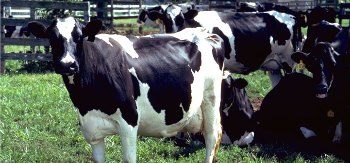
Mycoplasma mastitis is a threat to herd health.1,2 Mycoplasmas are highly contagious organisms that can infect the mammary gland and result in a severe case of mastitis that can be quickly transmitted to other cows causing significant herd outbreaks. A recent report indicated Mycoplasma bovis mastitis in cows was prevalent in California in the 1970s, in the Midwest by the 1990s, and by 2000 was a problem in Pennsylvania and New York. In 2003, 16 of 23 states involved in a herd survey had at least one dairy with a positive Mycoplasma culture from the bulk tank.
Mycoplasma spp. can also be present in both young and mature asymptomatic animals without clinical mastitis, and these animals can serve as carriers of the pathogen. Young animals can be exposed to Mycoplasmas at birth during calving from contact with the urogenital tract of an infected dam and in milk that they receive from shedding animals. In some herds, the infection can be manifested as ear infections in calves as well as pneumonia, arthritis, or a combination of both for animals of all ages. Cows in all stages of lactation are susceptible to the pathogen; however, based on field observations and a case report3, more cases are identified in cows following calving, when the stress of calving can impair immune function.
Please check this link first if you are interested in organic or specialty dairy production
Source
In many cases, the source of the pathogen is from purchased animals with unknown health status. Specifically, the primary sources of Mycoplasma species are infected udders, respiratory tracts, and urogenital tracts. Milk from all purchased animals should be cultured for Mycoplasma and other contagious mastitis pathogens before commingling newly acquired animals with the herd.
Signs and Symptoms of Mycoplasma Mastitis
- Cows have clinical mastitis in more than one quarter at a time.
- Milk samples from clinical cases of mastitis are negative on routine culture.
- Milk may appear watery, clotted, or tannish with sandy or flaky sediments that resemble cooked cereal in a whey-like fluid.
- An increase in the number of severe clinical cases of mastitis that resist antibiotic treatment.
- Milk production of affected cows decreases dramatically and, in severe cases, some cows may cease lactating.
- The mastitis outbreak was preceded by respiratory disease in bovines of different ages on the farm.
- An outbreak of ear infections or pneumonia in calves that resists antibiotic treatment preceded the mastitis outbreak.
- A severe lameness in animals, especially near calving.
Means of Spread
Mycoplasma spp. are spread from cow to cow during milking. Other sources include improper teat sanitation, contaminated intramammary treatments and treatment devices, milker’s hands, and airborne transmission in poorly ventilated barns. Mycoplasma mastitis can also result from spread of the organism from the respiratory or urogenital tract to the udder.
Basic Prevention and Control Measures
Maintain a closed herd. If animals are purchased, buy replacements from known Mycoplasma-free herds. Culture all replacements before commingling with the herd. Bulk tank samples may be cultured periodically to monitor for presence of Mycoplasma spp. in a herd. The presence of Mycoplasma spp. in the bulk tank sample almost always indicates that at least one cow in the herd has this disease. However, in herds that have cow(s) with Mycoplasma mastitis, approximately 25% of these cases will not be detected by bulk tank milk cultures. The reason is that sensitivity of bulk tank culture testing is reduced by cows with Mycoplasma mastitis that are shedding this pathogen into the milk at low numbers.
Sample Collection and Laboratory Analysis
Milk samples collected for culturing Mycoplasma spp. should be kept cool and plated within a few hours of collection to maximize isolation. If delay of more than four days is anticipated, samples should be frozen or stored in liquid nitrogen. It should be noted that freezing milk samples and storing samples frozen can greatly reduce the number of live Mycoplasma spp. Thus, such storage should be avoided when investigating a Mycoplasma mastitis problem.
The detection of Mycoplasmas in milk requires additional steps in the culture methods compared to the isolation of typical mastitis-causing pathogens. Therefore, if Mycoplasma mastitis is suspected, this should be communicated to the laboratory to ensure proper analyses are conducted to be able to identify the organism in the milk sample.
Steps to Take If Mycoplasma Is Identified in the Herd
No treatment is effective against Mycoplasma mastitis. Although there are rare reports of spontaneous recovery from Mycoplasma-infected cows, the infrequent occurrence of such cures dictates that infected animals should be removed from the herd.
If kept in the herd, infected cows should be segregated, double-identified with leg bands, and milked last or separately from uninfected cows. Do not have the infected cows in the same calving pens/groups that are used by uninfected cows.
Strict hygiene is essential! Use effective teat sanitizers. The milking system should be thoroughly sanitized following milking infected cows. Anyone coming in contact with the infected animal(s) should wear gloves and change gloves before touching an uninfected animal. Keep in mind that successful elimination of the pathogen from the herd usually includes culling of infected cows.
All fresh cows should be cultured for Mycoplasma.
Monitor success of elimination of the pathogen from the herd with weekly bulk tank analyses and periodic individual cow cultures of milk.
For more information, contact:
– Ms. Lynn Hinckley, Connecticut Mastitis Laboratory, (860) 486-4982, or
– Sheila M. Andrew, Ph.D., Extension Dairy Specialist, (860) 486-0803.
Fact sheet adapted from these sources:
1The NMC Newsletter “Udder Topics,” Mastitis Pathogen Notes: Mycoplasma species, 1999.
2 Ruben Gonzalez. 2005. Mycoplasma Mastitis – What Is New in the Northeast? Proc. of the National Mastitis Annual Regional Meeting, Burlington, Vermont.
3 Wilson, et al., 2007. J. Am. Vet. Med. Assoc. 230:1519.
Author
Sheila M. Andrew
University of Connecticut